

Women’s Well Scan

So many women are concerned with breast disease, as they should be. However, inflammatory cardiovascular disease is the primary cause of death and degeneration here in America.
Well Scan Full Body - Women's Assessment includes:
Cardiovascular / inflammation patterns in the arteries and lymphatic system throughout the body
Stress response / hormone tissue inflammatory patterns
Digestive inflammation patterns
Joint and soft tissue inflammation
When these systems are working optimally with little inflammation the body feels like a well orchestrated symphony playing the the tune of Well Being.

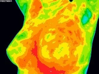


These are thermography photos of a client, three months apart. The reddish color indicates a lot of inflammation and congestion in the tissues. This is an alarming report indicating unfavorable tissue changes. The client was under extreme stress, her symptoms included menstrual pain, tender breast tissue, digestive bloating and gas. After a 21 day purification, patient reports no more gas and bloating, and PMS symptoms are minimal. The Patient does bi-annual purification to maintain.
Breast Thermography is an essential addition to breast health screening because of its ability to monitor physiological changes in the tissue, such as hormone imbalances and ramifications of a person’s lifestyle choices. These scans are performed with ZERO adverse affects to the body.
Many women face conditions such as fibrocystic breast tissue, estrogen dominance and Polycystic Ovarian Syndrome (PCOS). This is visually seen on the scan and reported by the radiologist. From these findings, a unique protocol of dietary changes, removal of endocrine disruptors in the home and environment, and whole food supplements that feed and nourish the female endocrine system may be suggested. Simple changes create profound improvements resulting in a balanced, healthy endocrine system.
If the report is abnormal or shows that a hormone imbalance is affecting the breast tissue, additional diagnostic imaging may be suggested.


These are thermography photos of a client taken at the initial visit, twelve months later, twenty four months later, and twenty seven months. The warmer areas indicates the progression of hormone imbalance getting more inflamed at each scan.


These are thermography photos of a client with a progressing breast cancer diagnosis. The red and orange areas show the progression in the right breast.

Dr. William Hobbins is a former surgeon who pioneered breast cancer detection through both mammography and thermography. Now 90, he continues to urge widespread use of thermography for initial screening and prevention because “the angiogenesis of a breast cancer is not only the earliest sign, but the greatest sign for detection and prognosis in treatment.”